FOURTEEN ORDERS OF MAGNITUDE
From Planet to Cell
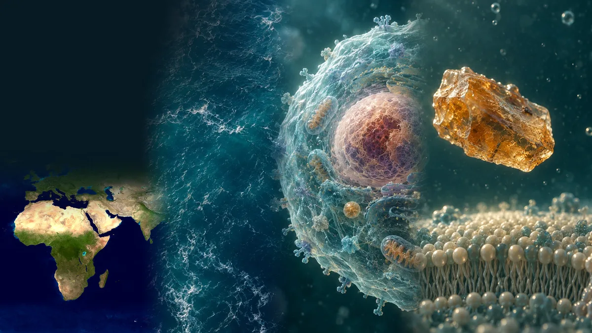
A 22-second animated descent from the whole Earth to a particle 100 nm across, in six stops. Each stop gets one real reference size, from the planet's 12,742 km diameter to a cell membrane a few nanometers thick. The frames are illustrations of scale, not microscope images.
 Illustration
The route: one planet, six stops, 22 seconds
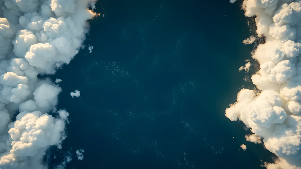
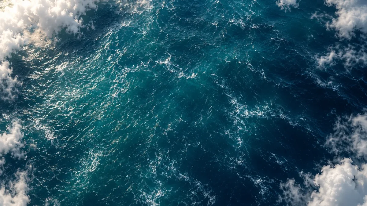
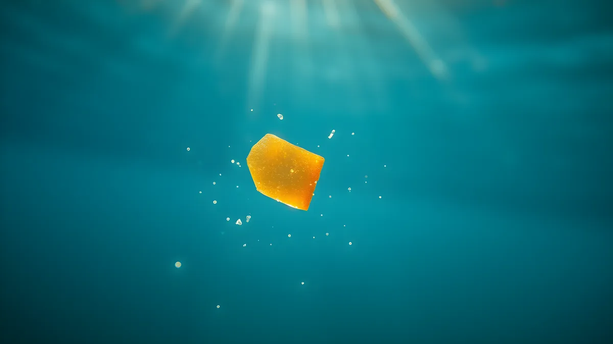
"From planet to cell" opens from the rotating Earth on the home page and runs for just under 22 seconds at normal speed. It stops six times: the planet, the atmosphere, the water just below the surface, a millimeter of seawater, a cell and one particle 100 nm across. The journey can be paused, scrubbed or jumped stop by stop.
The scale bar falls from 10,000 km to 100 nm, a span of 14 orders of magnitude (level P; our calculation: ten million meters to one ten-millionth of a meter). The last particle is smaller than the shortest wavelength the human eye detects, 380 nm1. A conventional light microscope cannot resolve its shape, because Abbe's limit for such instruments is about 0.2 µm2.
14
orders of magnitude from the first to the last stop
journey scale bar, 10,000 km to 100 nm (10⁷ m to 10⁻⁷ m); our calculation3
P: arithmetic on the fixed chapter scales, not a scientific finding; Earth's 12,742 km diameter gives the same order
PProvenance≈12,742 km
Earth's mean diameter
twice NASA's volumetric mean radius of 6,371 km (doubling is our calculation)3
A: official NASA planetary reference value
AConsensus≈0.2 µm
resolution limit of a conventional light microscope
Abbe's diffraction limit, about half the wavelength of light; super-resolution fluorescence methods bypass it for labeled molecules2
A: textbook optics, stated by the Nobel committee; a smaller particle can still show up as a blur, but its shape is not resolved
AConsensusThe scale ladder from 10,000 km to 100 nm
Each stop is a power of ten. One real object anchors each one, and the factor to the next stop shows how far the camera moves.
- 10,000 km, planet. Earth's mean diameter is about 12,742 km, twice NASA's mean radius of 6,371 km3. Next stop: 10,000 times closer.
- 1 km, atmosphere. Low clouds, the kind the camera passes through here, have their bases between the surface and about 2 km in the mid-latitudes (level B)4. Next: 1,000 times closer.
- 1 m, water. The 1 m on the scale bar describes the frame, not a depth: the camera is just below the surface. Little sunlight reaches below 200 m, the lower edge NOAA gives for the sunlit (euphotic) zone5, and the ocean averages 3,682 m deep6. Next: 1,000 times.
- 1 mm, microworld. An average human hair is about 70 µm thick7; about 14 would fit side by side across 1 mm (level P; our calculation). Next: 100 times.
- 10 µm, cell. A human red blood cell is 7–8 µm across8; a typical cloud droplet is about 20 µm (level B)9. An E. coli bacterium, about 1 µm wide and 2 µm long8, sits between this stop and the next. Next: 100 times.
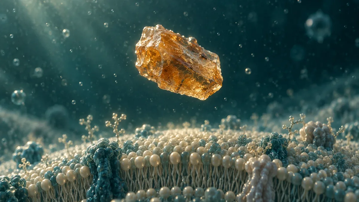
- 100 nm, nanoparticle. Visible light spans 380–700 nm1. A cell membrane's lipid bilayer is about 4 nm thick, and the whole membrane with its proteins is reported at 4–10 nm8. A 100 nm particle is therefore 10 to 25 times thicker than the membrane beside it (level P; our calculation).
From planet to cell: six stops and reference sizes on one logarithmic ruler
Data table
| label | value | unit | kind | source | note |
|---|---|---|---|---|---|
| Stop 1 · Planet (journey scale) | 10000000 | m | stop | site-original | editorial stop fixed by the journey code |
| Earth, mean diameter | 12742000 | m | reference | S-300 | |
| Ocean, average depth | 3682 | m | reference | S-301 | |
| Low clouds, base up to about 2 km | 2000 | m | reference | S-306 | mid-latitudes; draw as a band from the surface to 2.0e3 |
| Stop 2 · Atmosphere (journey scale) | 1000 | m | stop | site-original | editorial stop fixed by the journey code |
| Sunlit (euphotic) zone, lower edge | 200 | m | reference | S-302 | |
| Stop 3 · Water (journey scale) | 1 | m | stop | site-original | editorial stop fixed by the journey code |
| Stop 4 · Microworld (journey scale) | 0.001 | m | stop | site-original | editorial stop fixed by the journey code |
| Human hair, average diameter | 0.00007 | m | reference | S-231 | |
| Cloud droplet, typical diameter | 0.00002 | m | reference | S-303 | |
| Stop 5 · Cell (journey scale) | 0.00001 | m | stop | site-original | editorial stop fixed by the journey code |
| Red blood cell, diameter 7–8 µm | 0.0000075 | m | reference | S-222 | |
| E. coli, length | 0.000002 | m | reference | S-222 | |
| Visible light, 380–700 nm | 5.4e-7 | m | reference | S-304 | draw as a band from 3.8e-7 to 7.0e-7 |
| Light microscope limit (Abbe) | 2e-7 | m | reference | S-305 | |
| Stop 6 · Nanoparticle (journey scale) | 1e-7 | m | stop | site-original | editorial stop fixed by the journey code |
| Cell membrane, 4–10 nm | 4e-9 | m | reference | S-222 | draw as a band from 4e-9 to 1.0e-8 |
What the journey is not
The opening Earth is the NASA Blue Marble globe; the ocean, cell and particle frames are AI illustrations, not photographs or microscope images. The camera path is one continuous illustrative move; no single particle was tracked from sea to cell.
The final frame is not drawn to scale: a real 100 nm particle would be 10 to 25 times thicker than the membrane, not a few times. Nor is it evidence that particles enter cells; what is known about uptake is on Human Body. Size classes are on What Is Nanoplastic, detection on How We Measure It, the panels on Four Microworlds.
 Illustration
 Illustration
 Illustration
 Illustration
Sources
- NASA Science (2023). Visible Light. NASA. science.nasa.gov/ems/09_visiblelight/ web open access
- The Royal Swedish Academy of Sciences (2014). The Nobel Prize in Chemistry 2014: popular information (How the optical microscope became a nanoscope). Nobel Prize Outreach / The Royal Swedish Academy of Sciences. www.nobelprize.org/prizes/chemistry/2014/popular-information/ press open access
- Williams DR (2024). Earth Fact Sheet. NASA Goddard Space Flight Center, NSSDCA. nssdc.gsfc.nasa.gov/planetary/factsheet/earthfact.html dataset open access
- UCAR Center for Science Education (2026). Cloud Types. University Corporation for Atmospheric Research (UCAR). scied.ucar.edu/learning-zone/clouds/cloud-types web open access
- NOAA National Ocean Service (2026). How far does light travel in the ocean?. National Oceanic and Atmospheric Administration (NOAA). oceanservice.noaa.gov/facts/light_travel.html web open access
- NOAA National Ocean Service (2026). How deep is the ocean?. National Oceanic and Atmospheric Administration (NOAA). oceanservice.noaa.gov/facts/oceandepth.html web open access
- US Environmental Protection Agency (2026). Particulate Matter (PM) Basics. US EPA, last updated 18 September 2026. www.epa.gov/pm-pollution/particulate-matter-pm-basics web open access
- Milo R, Phillips R (2015). Cell Biology by the Numbers. Garland Science. doi:10.1201/9780429258770 report open access
- Russell R (2007). Sizes of Aerosols, Raindrop and Cloud Droplets. UCAR Center for Science Education. scied.ucar.edu/image/aerosols-raindrop-cloud-droplets-sizes web open access
Evidence levels and provenance
- AConsensusUsed on this page
- Multiple independent studies or a systematic review plus a statement by a scientific body.
- BWell supportedUsed on this page
- Several independent studies with concordant findings.
- CLimited
- Few studies, narrow conditions, or methodological caveats.
- DDisputed
- Conflicting results; both sides are shown.
- EUnknown
- No reliable evidence found; the search scope is stated.
- PProvenanceUsed on this page
- Provenance: checked against the primary record. A separate category, not an evidence grade: used for bibliographic facts, legal texts, official metadata, definitions quoted from a standard, the site's own arithmetic and statements about the site's process.
Editorial categories of this site. A–E grade scientific findings, per claim, with a one-line rationale; D and E are not lower rungs of the A–C scale. P is not a grade: it marks facts checked against their primary record.